
PixelFold


A protein structure viewer and residue interaction network engine that runs entirely in your terminal.
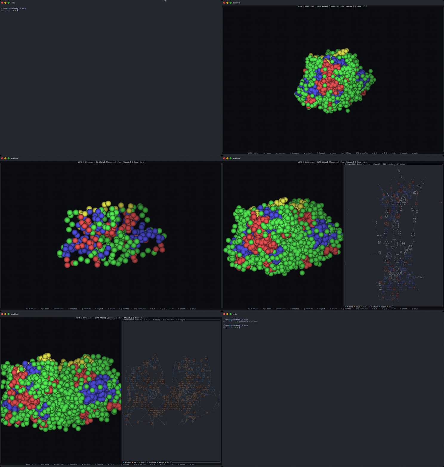
Validation
The viewer is the demo. The interaction engine is the product, and it is measured against existing trusted tools.
Validated 205 benchmark entries against DSSP 4, FreeSASA, PLIP and RING.
| Metric | Reference | Entries | Result |
|---|---|---|---|
| Secondary structure (Q3) | DSSP 4 | 194 | 97.1% |
| Backbone H-bond edges (F1) | DSSP 4 | 205 | 0.97 |
| SASA mean abs error | FreeSASA | 203 | 4.77 A^2 |
| SASA median abs error | FreeSASA | 203 | 3.61 A^2 |
| SASA correlation (Pearson r) | FreeSASA | 203 | 0.99 |
Per-type interaction agreement (precision / recall / F1):
| Interaction type | Reference | Entries | Pred edges | Ref edges | Precision | Recall | F1 |
|---|---|---|---|---|---|---|---|
| hydrogen-bond | PLIP | 60 | 676 | 519 | 0.64 | 0.82 | 0.68 |
| salt-bridge | PLIP | 60 | 129 | 127 | 0.91 | 0.93 | 0.87 |
| hydrophobic | PLIP | 60 | 435 | 377 | 0.87 | 0.96 | 0.90 |
| pi-stacking | PLIP | 60 | 23 | 22 | 0.97 | 0.95 | 0.92 |
| pi-cation | PLIP | 60 | 6 | 17 | 0.97 | 0.88 | 0.84 |
| halogen-bond | PLIP | 60 | 4 | 5 | 1.00 | 0.98 | 0.98 |
| water-bridge | PLIP | 60 | 511 | 345 | 0.59 | 0.83 | 0.65 |
| metal-coordination | PLIP | 60 | 444 | 338 | 0.82 | 0.92 | 0.81 |
| hydrogen-bond | RING | 205 | 49374 | 43512 | 0.79 | 0.90 | 0.83 |
| salt-bridge | RING | 205 | 4251 | 1545 | 0.38 | 1.00 | 0.50 |
| pi-stacking | RING | 205 | 517 | 3756 | 1.00 | 0.25 | 0.32 |
| pi-cation | RING | 205 | 453 | 454 | 0.72 | 0.71 | 0.62 |
| pi-hydrogen-bond | RING | 205 | 269 | 97 | 0.65 | 0.99 | 0.66 |
| halogen-bond | RING | 205 | 7 | 9 | 0.99 | 0.98 | 0.97 |
| metal-coordination | RING | 205 | 567 | 558 | 0.89 | 0.91 | 0.86 |
| disulfide | RING | 205 | 249 | 249 | 1.00 | 1.00 | 1.00 |
These are agreement figures and never improvement claims. Where PixelFold and the reference disagree, the reference is right by construction. The harness lives in this repository and runs in CI, allowing the numbers above to be reproduced, rather than simply asserted. See Validation and methods for what the weaker rows mean.
Install
This needs Rust 1.88 or newer. Each GitHub release also carries prebuilt binaries for macOS, Linux and Windows, with shell and PowerShell installers. From v2.1.0 the Linux binaries need glibc 2.28 or newer, which covers RHEL/Rocky 8 and later, the usual cluster login node; earlier ones need glibc 2.34.
A 30 second demo
Dihydrofolate reductase with its inhibitor methotrexate. How does this drug interact with the enzyme, and does it matter structurally?
$ pixelfold interactions 4DFR --ligand MTX
kind           chain_a  resi_a  resn_a  atoms_a     chain_b  resi_b  resn_b  atoms_b  distance  angle
-------------  -------  ------  ------  ----------  -------  ------  ------  -------  --------  -----
hydrogen-bond  A            52  ARG     NH2         A           161  MTX     O            2.88  152.8
hydrogen-bond  A            52  ARG     NH2         A           161  MTX     OE2          3.43  138.2
hydrogen-bond  A            57  ARG     NH1         A           161  MTX     O1           2.57  155.1
hydrogen-bond  A            57  ARG     NH2         A           161  MTX     O2           2.72  170.8
... 48 rows
Next, analyze the network to find where the important residues are located.
$ pixelfold rin 4DFR --analyze
Residue interaction network: 311 residues, 615 edges, 5 components
...
Top hubs by betweenness centrality:
  residue    resn  ss  degree      between
  A/23       ASN   C        5     14016.66
  A/24       LEU   C        4     13113.96
  A/161      MTX   C       15     11017.94
  B/162      MTX   C       17     10686.73
  B/22       TRP   C        7     10301.22
Both copies of the inhibitor rank among the most central residues in the structure. A/161 is also an articulation point, where the network will split if it is removed. This local computation takes about a second, and provides insight to the fold.
Everything here is scriptable. --format json|tsv|graphml feeds a pipeline,
--select narrows any command with the same query language, and pixelfold 4DFR
opens the interactive viewer over the same data.
Why it exists
Structures usually live where the compute is, like a cluster login node, a remote workstation, or a container. To look at one normally, one must copy it to their machine, open a desktop application, before any analysis can be made.
PixelFold runs in the shell that already has the file, with no need for X11, a display server,
a GPU, browser upload, and Python environment. It is a
single static binary, which drops into a Nextflow or Snakemake step as easily as
grep. The analysis is in text, so it diffs, greps, and pipes, while being
deterministic.
Honest comparison
PixelFold is is in its infancy. It is not competitive on rendering, breadth, or maturity with any established viewer, and it does not claim to be the reference on interaction chemistry. Below is a compilation of how different tools compare.
| Tool | Where it beats PixelFold | Where PixelFold differs |
|---|---|---|
| PyMOL | Ray-traced publication figures, cartoons, surfaces, electrostatics, trajectories, twenty years of plugins, an embedded Python API, and near-universal adoption | Terminal-native with no display server; MIT throughout; the network is the primary output rather than a side feature |
| Mol* | GPU rendering at a quality and scale PixelFold cannot approach, million-atom and integrative models, MD streaming, zero install, and it powers RCSB, PDBe and AlphaFold DB | No browser and no upload, so embargoed coordinates stays local. PixelFold complements through its exportable .mvsj scenes |
| ChimeraX | Cryo-EM density, structure building and refinement, VR, AlphaFold workflows, a bundle ecosystem, and NIH-funded maintenance | MIT versus a non-commercial licence, so it can be vendored freely; a small static binary rather than a GB-class install |
| RING 4.0 | The peer-reviewed network reference the field accepts. Ligand coverage across the whole PDB chemical component dictionary. RING-PyMOL already does ensemble and MD contact analysis | Readable MIT source you can fork or embed as a Rust library, offline with no web upload, and graph analytics in the same binary. Distribution differences, not accuracy claims |
| PLIP | The yardstick for interaction detection. Its protonation-aware pipeline is more physically grounded, and it covers DNA, RNA and protein-protein interfaces | No Python or OpenBabel to install; profiles the whole structure as a graph rather than only ligand sites |
| asciiMol | Runs on genuinely any terminal, including plain ncurses, where PixelFold degrades to half-blocks and looks worse. pip install into an environment you already have. Native .xyz and SMILES |
Protein-scale mmCIF and PDB with chains, conformers and a selection language, and the analysis engine behind it |
Validation and methods
Reference implementations: DSSP 4 for secondary structure, FreeSASA for
solvent-accessible surface area, PLIP for the interactions of bound ligands, and
RING 4.0 for interactions across the whole structure, over a stratified benchmark
of 205 PDB entries, 60 of them protein-ligand complexes. Run it yourself with
cargo run -p pixelfold-validate -- --check.
Each interaction reference is scored only over the part of the structure it looks at. Against PLIP, only PixelFold's edges that touch a ligand PLIP profiled count. Against RING, edges to water are left out, as are pairs of amino acids fewer than three residues apart in one chain, which RING never pairs at its defaults.
Interaction geometry follows PLIP's published thresholds, and RING's for the
pi-hydrogen bond, which PLIP does not detect, with the exact source recorded
next to every constant in
params.rs. Each known
difference from RING is also noted. Secondary structure is
Kabsch-Sander electrostatic hydrogen bonding with inferred amide hydrogens.
The weak rows are a modelling choice, and they are the honest headline. Hydrogen bonds (F1 0.68, precision 0.64) and water bridges (F1 0.65, precision 0.59) are the two classes where PixelFold reports what the geometry allows while PLIP commits to one reading of it. Of the 261 hydrogen-bonded residue pairs PixelFold finds and PLIP does not, 94 come from a rotatable hydroxyl, which PixelFold turns toward each acceptor and PLIP fixes wherever OpenBabel placed its hydrogen; 77 also form a salt bridge, which PLIP then drops and RING keeps; and the other 90 have nitrogen donors, many of them histidine tautomers, amide flips and ligand nitrogens the coordinates leave open. A water bridges up to two donors and two acceptors, one per lone pair and per hydrogen, where PLIP keeps one donor per acceptor and drops a donor that already hydrogen-bonds the ligand. Those two classes are roughly half of all protein-ligand interactions, so for ligand-site work where a curated answer matters, use PLIP.
Against RING, the weak rows come from following PLIP's definitions. RING 4.0 draws three types differently, and where the two disagree RING is the reference. Salt bridges (F1 0.50, precision 0.38) are a superset: PixelFold finds 1541 of RING's 1545 and 2710 more, because PLIP pairs charge centres up to 5.5 A apart where RING asks for about 4.0 A between atoms, and RING pairs only amino acids. Pi-stacking (F1 0.32, recall 0.25) runs the other way: 515 of PixelFold's 517 stacks are in RING, but RING's wider window (about 6.5 A between ring centres, against PLIP's 5.5 A with at most 2.0 A of lateral offset) finds seven times as many. Pi-cation (F1 0.62) disagrees in both directions: PixelFold, like PLIP, counts histidine as a cation and needs the cation within 2.0 A of the ring's axis, while RING's cations here are only arginine and lysine, within about 5.0 A. Hydrogen bonds agree at F1 0.83 (precision 0.79): RING's shorter 3.9 A cutoff and its added hydrogen-to-acceptor limit leave it fewer bonds. Pi-hydrogen bonds (F1 0.66, precision 0.65) follow RING's own definition, and PixelFold finds 95 of RING's 97 and 174 more. Of those, 97 come from a rotatable hydroxyl or ammonium, which PixelFold turns toward the ring and RING fixes at one torsion; the other 77 pass RING's published criteria and are still absent from its server's output, for reasons its closed engine does not show. The halogen-bond rows rest on five and nine reference edges, and an entry where neither side finds one counts as agreement, so they say little.
Per-edge interaction energies are a literature-sourced scoring table with one
primary citation per value, in
energy.rs. They are
representative magnitudes, not computed free energies.
Architecture
A Cargo workspace. pixelfold-core has no terminal or rendering dependencies, so
the analysis is usable as a library.
pixelfold-core: parsing, DSSP, SASA, the interaction engine, the selection language, the residue interaction network with its graph analytics, and ligand pose comparisonpixelfold-render: a z-buffered sphere-impostor rasteriser writing one RGBA framebuffer, and the sinks that put it on a terminal (Kitty, iTerm2, truecolor half-blocks) or in a PNGpixelfold-tui: theratatuifront end and the linked network panepixelfold-cli: thepixelfoldbinarypixelfold-validate: the agreement harness
The CLI computes everything the viewer shows headlessly.
Limitations
Being explicit about what this does not do:
- The RING reference comes from its web server. RING's standalone binary is licensed to one person and cannot be passed on, so the RING golden files were generated by the public RING 4.0 web server, which reports no version. The committed files keep the comparison reproducible offline, but regenerating them depends on that server, and its anonymous rate limit makes a full rerun take about a day and a half.
- Hydrogen bonds and water bridges over-report relative to PLIP, as above.
- Salt bridges over-report and pi-stacking under-reports relative to RING, as above, because both follow PLIP's geometry. Pi-hydrogen bonds over-report relative to RING too, mostly from rotatable hydrogens.
- No cartoons or ribbons, no molecular surfaces, no ray tracing. Space-filling spheres and bonds only.
- Poses are matched to the reference by sequence, not by structure.
pixelfold posespairs each pose chain with the reference chain it aligns with, so a co-folding model numbered from 1 compares with a crystal structure in author numbering. Of identical chains, as in a homodimer, the pairing that keeps the most reference contacts wins; a cofactor or an ion matches the reference residue of the same component. A chain must share at least half its residues with its partner (a peptide must match whole), so a pose of another protein is refused, and a contact at a substituted residue counts when its kind agrees. A reference holding several copies of the ligand needs one picked with--reference-select. When the reference ligand is one covalent piece, a pose ligand in several pieces that each hold its residues, or its heavy atoms by element, holds copies of it: the pose is scored by the copy most like the reference's (the most similar, then the nearest), named in thecopycolumn. Pieces that are not all copies are scored as one, with a warning. A pose file without its ligand's chemical definitions, as co-folding output often is, borrows the reference's by atom name; a ligand named differently in the two gets a warning that its contacts are incomplete. An SD file's atoms carry no names, so a docked ligand takes the reference's names, and with them its chemistry, by pairing the two bond graphs, bond orders included where they agree; a ligand whose bond orders agree in no pairing, as another tautomer's would not, is paired by its bonds alone with a warning, and a molecule other than the reference's ligand is scored untyped, with a warning. Only V2000 SD files are read, and a docked ligand's charges are not used. Water-mediated contacts are left out, since predicted poses carry no water. The ligand RMSD is AlphaFold 3's pocket-aligned one, symmetry-corrected and indifferent to the order of a ligand's copies; a pose whose ligand atoms pair with the reference's neither by name nor by bond graph (a ligand missing atoms, or one renamed and so distorted its bonds no longer read) gets none, with a warning. The same pairing names the reference ligand's tetrahedral stereocentres a pose inverts (inverted, empty like the RMSD when the atoms cannot be paired); a centre the pose leaves flat, as a covalent warhead without its bond to the protein is, does not count. The bond graph is heavy-atom connectivity without bond orders, so a centre whose substituents differ only in bond order or hydrogens (glyceraldehyde's CHO beside its CH2OH) is not checked, nor is one whose neighbours a symmetry could exchange (cis and trans across a symmetric ring, as in 4-methylcyclohexanol), one left with fewer than three neighbours inside the ligand selection (a glycan's anomeric carbon on its asparagine).invertedalso names the reference's E and Z double bonds a pose turns the other way (GPP A/101 C2=C3), read from its chemical definitions: a double bond between carbons or nitrogens outside any ring of fewer than eight atoms, with a substituent on each end that no symmetry exchanges. Thegeometrycolumn holds PoseBusters' checks of the pose's own geometry, with the reference ligand's geometry in place of RDKit's ideal one and PoseBusters' tolerances around it: bond lengths and angles more than a quarter off, atoms neither bonded nor sharing a neighbour within seven tenths of their lower bound (never further apart than the reference holds them, so a metal the bond graph leaves unbonded is no clash), and aromatic rings and carbon double bonds more than 0.25 A from flat where the reference's are flat. A strained reference ligand so passes its strain on, PoseBusters' energy ratio is not computed, and without the reference ligand's definitions neither flatness nor E and Z is checked. A pose whose atoms are named otherwise is paired by bond graph alone, which cannot tell a double bond from a single one that a symmetry of the graph exchanges it with, so such a double bond and its flatness go unchecked; and one distorted past reading its bonds pairs not at all, so it gets no checks, only the warning that it has no RMSD. - No trajectories. A multi-model file such as an NMR ensemble works model
by model (
--model N, or--model allfor a report on every model and one interaction network over them all), but a molecular-dynamics trajectory is not read; frames written as the models of one file are, andrinfolds separate files, such as a co-folding model's samples, into one network. - No cryo-EM density, no electrostatics, no structure building or refinement.
- Rendering quality depends on your terminal. Kitty and iTerm2 show real
pixels; anywhere else falls back to half-blocks, which is coarse. Pass
--protocol half-blockif the structure pane comes up blank. - Not peer reviewed. The validation harness is the evidence on offer.
Usage
# Interactive viewer, from a file or a 4-character PDB id (fetched and cached)
# Non-covalent interactions
# The residue interaction network, for Cytoscape, Gephi, or a script
# Ligand poses, from co-folding, docking or an NMR ensemble, ranked by the
# reference contacts each keeps, with their pocket-aligned RMSD, their clashes,
# the stereocentres and double bonds they invert, and what of their own
# geometry strays from the reference ligand's;
# every model is a pose, and residues are matched by sequence, so the numbering
# may differ
# One copy of a ligand the reference holds several of; a pose holding several
# is scored by the copy most like the reference's
# The same ranking as a table in the viewer, each pose drawn on the reference's
# pocket
# Docked ligands from SD files, each record a pose, laid into the reference
# without its ligand, or into the receptor they were docked into
# Secondary structure and solvent accessibility
# Render to a PNG, or to the terminal
# One model of an NMR ensemble, or every model with a model column
# One network over every model, each edge weighted by the share of models it
# is in; the edges in at least nine models in ten, analysed
# The same over a prediction's samples, each its own file; they must number
# residues alike
# The biological assembly instead of the deposited coordinates; copies of a
# chain are named A, A-2, A-3
# Any command, narrowed by the selection language
Viewer controls
| Key | Action |
|---|---|
WASD |
Rotate; z/x roll |
+/-, arrows |
Zoom, pan |
1/2 |
All atoms, C-alpha backbone |
c b h |
Bonds, B-factor colouring, hydrogen bonds |
i, click |
Inspect mode, pick an atom; r highlights its residue |
g |
Linked interaction network pane |
l e |
Network layout (force or spatial), node colour (structure or burial) |
t y |
Filter network edges by interaction type, by chain |
k [ ] , . |
Slab: toggle, move, and resize a depth slice |
m/M |
Next, previous model of a multi-model file, or pose of the pose table |
o p |
Pose table (poses --view): sort by the next column, hide or show |
n |
Network of the whole ensemble, edges brightened by the share of models they are in; again for those in half, then nearly all, of the models; again for the shown model's |
u |
Biological assembly on or off |
f, q |
Reset the view, quit |
Citations